



Contents
Scroll to:
https://doi.org/10.23947/2541-9129-2026-10-1-47-60
EDN: BZBQLB
Scroll to:
Introduction. Fatigue failure is one of the main causes of failure of metal structures subjected to variable loads. Initially, this damage is not visible as cracks, but it leads to the accumulation of microdefects and the redistribution of internal stresses. Currently, it is not possible to monitor the progression of these defects in large structures with a significant surface area. To detect such processes in a timely manner, highly sensitive inspection methods are required that can identify potential areas of failure with a high degree of accuracy during the early stages of structural operation. Such methods do not currently exist, and our research aims to solve this problem to a certain extent. One promising approach is the monitoring of changes in the strength of a permanent magnetic field, which reflects the evolution of material state. The current study aims to investigate the potential of spatial analysis of magnetic response to identify instability zones during fatigue loading, where the likelihood of failure is high, as well as to analyze changes in steel structure.
Materials and Methods. The study focused on samples made of 09G2S steel, subjected to loading to fracture on a servohydraulic testing machine INSTRON-8801. Magnetic measurements were taken at 12 points along the sample using an IKN-2M-8 instrument. Changes in the resulting strength of the permanent magnetic field were recorded at different stages of fatigue loading. All measurements were repeated at least three times to ensure the reliability of the results.
Results. It has been found, that at the stage of relative operating time Ni/Np = 0.4–0.5, anomalous changes in the magnetic field strength corresponding to the fracture nucleus were recorded at certain points. Additionally, a characteristic area of signal stabilization was observed in the range Ni/Np = 0.8–0.9. This could be explained by the temporary relaxation of stresses prior to destruction. The obtained data demonstrate the local variability of the magnetic response and confirm the sensitivity of this method to the early stages of material degradation.
Discussion. The conducted research has shown that spatial analysis of changes in the strength of a permanent magnetic field can be used to locate fracture nuclei in ferromagnetic steels. This dataset can be used as a basis for training samples for intelligent monitoring systems, including neural network algorithms that focus on predicting the remaining life and automatically assessing the technical condition of structures. This is particularly important for welded structures with a high number of welds.
Conclusion. The introduction of energy into a system inevitably leads to a reorganization of the structure of the material in order to adapt to external forces. This reorganization is accompanied by a change in the material's magnetic field. By recording these changes, it is possible to interpret the measurement results in terms of possible destruction, as the most efficient way for the system to utilize the supplied energy is through the formation of new surfaces, or cracks.
Shermatov D.N., Borisov A.O., Gafarova V.A., Kuzeev I.R. Local Gradient Indicator of Magnetic Variability under Cyclic Loading of Steels. Safety of Technogenic and Natural Systems. 2026;10(1):47-60. https://doi.org/10.23947/2541-9129-2026-10-1-47-60. EDN: BZBQLB
Introduction. The importance of accurately assessing the remaining lifespan of metal structures stems from the need to balance industrial safety with operational costs. This issue is particularly relevant for extended welded shells, tanks, and pipelines, which require monitoring a large surface area and substantial amounts of metal under conditions of limited access and challenging loading conditions.
Traditional methods of non-destructive testing, such as ultrasound, radiography, and acoustic emission, are effective at detecting formed defects like macrocracks and discontinuities. However, these methods have limitations when it comes to an early detection of fatigue damage. They usually require careful surface preparation, significant labor costs, and are not well-suited for rapid mapping of extended welds. Additionally, they often provide an integrated assessment of the condition, without the ability to locate the initial areas of degradation or residual stress gradients. As a result, a significant portion of the design effort is spent blindly, potentially leaving dangerous areas undetected until the stage when macrocracks have developed.
In this context, here has been a growing interest in methods that rely on recording the material's own physical fields. These methods are sensitive to changes in the internal state of the material without directly injecting energy into the controlled volume. One of the most promising methods is the analysis of the intensity distribution of the permanent (residual) magnetic field that forms in ferromagnetic steels as damage accumulates and residual stresses redistribute. Magnetic methods have a number of important advantages: the possibility of remote measurements, high speed of examination of large areas, and sensitivity to the early stages of stress and defect redistribution, when macrocracks have not yet formed [1].
Reliable diagnosis of the remaining life of metal structures relies on a thorough analysis of microscopic changes that occur in the material's structure as fatigue damage accumulates. One of the most sensitive and informative indicators of these changes is the material's magnetic field, which forms as a result of changes in its domain structure. Changes in the domain configuration integrally reflect a combination of microstructural transformations and the evolution of residual stresses, making them a valuable basis for developing highly accurate diagnostic criteria.
In ferromagnetic materials, there are significant differences in the magnetic domain structure near the surface compared to the interior. Experimental studies have shown that domain walls near the surface can be significantly wider than inside the material. This leads to a decrease in magnetic energy density in the wall and, as a result, affects the mobility of domain boundaries and their response to local mechanical stresses and defects [2]. Therefore, it is important to take into account the special magnetic properties of surface layers when analyzing the overall magnetic texture of a material.
Direct visualization of internal domain structures within the volume of solid materials is still a challenging task due to the limited spatial resolution of most magnetic imaging techniques. Techniques such as magneto-optical imaging and magnetic force microscopy can only effectively record magnetic morphology on the surface of a sample. Instead, indirect approaches are used to study the three-dimensional magnetic structure. Classical micromagnetic models and multiscale numerical simulations allow us to predict domain configurations and their dynamics by taking into account both the local magnetic fields and the crystallographic properties of the material. For example, the introduction of dislocation stresses into micromagnetic simulation demonstrates how dislocations can serve as anchor points for domain boundaries and influence the Barkhausen effect [2].
It is worth mentioning the latest methods for reconstructing magnetic structures based on machine learning. In [3], a model based on a convolutional neural network (MagNet) is presented, which increases the accuracy of reconstructing the three-dimensional configuration of magnetization from tomography data. This approach overcomes the limitations of classical vector tomography algorithms, eliminating the artifacts of incomplete data and significantly improving the quality of the reconstructed magnetic field.
A similar principle was applied in the study [4], where a neural network model is trained to transform images of an external magnetic field (for example, leakage field maps) into the distribution of the magnetization vector inside the material. This makes it possible to reconstruct complex domain textures with a variable direction of magnetization, which are inaccessible to traditional inversion methods [4]. Such neural network approaches expand the possibilities of interpreting experimental data and bring researchers closer to the direct reconstruction of the internal magnetic texture by indirect measurements.
The microstructure of the material, including the distribution of magnetization, changes as a result of plastic deformation under cyclic loading conditions. Modern measurement methods do not directly track the movement of individual magnetic domains within a material, but the cumulative magnetic response, such as a hysteresis loop, is sensitive to these microstructural changes. The restructuring and reorganization of domain boundaries on a microscopic level is manifested in measurable changes in magnetic permeability, coercive force, and other material parameters. Thus, the analysis of the dynamics of magnetic characteristics under load makes it possible to judge changes in the internal structure. By taking these effects into account, we can move from an overall assessment of the material's condition to a more localized analysis aimed at detecting areas that are prone to damage.
The internal (volumetric) behavior of the material during fatigue deformation differs significantly from the processes occurring near the surface. In the thickness of the material, plastic deformation is distributed more evenly and the gradients of residual stresses are significantly lower than at the surface [5]. In a polycrystalline volume, dislocations are generated and accumulate in groups, causing significant local stresses, which are difficult to remove due to the lack of a free surface. Grain boundaries act as an internal "surface", however, for dislocations to escape through these boundaries, an energy barrier must be overcome [6]. Only after reaching a critical level of accumulated stresses and defect energy [7], it is possible for microcracks to form inside the material. These features are consistent with the Mura concept of relaxation of internal stress fields [8], as well as with experimental data on the uneven distribution of residual stresses (stress anisotropy) in steels [9].
Unlike the volume, the free surface of the material acts as an effective source for dislocations, requiring significantly less energy for defects to form. This explains why fatigue cracks often initiate on the surface. As a result, changes occurring in the subsurface layer during loading can serve as informative diagnostic indicators of developing degradation.
One of these signs is the formation of self-similar (fractal) structures on the surface as defects develop. These structures have the property of self-similarity at different scales, as noted by B. Mandelbrot [10]. The morphology of the damaged metal surface often exhibits fractal characteristics, which can be quantified [11]. The analysis of relief elements at various scales helps to establish a connection between surface fatigue and volume destruction processes [12].
The heterogeneity and roughness of the surface can also affect the magnetic properties of the subsurface layer. Research has shown that changes in the fractal structure of a deformed material correlate with its magnetic parameters, such as saturated magnetization and magnetic permeability [11]. In other words, as damage increases, the distribution of physical properties may exhibit multifractal features. Tracking the evolution of several such multifractal parameters expands the possibilities of diagnostic interpretation, allowing for more reliable identification of the early stages of material degradation.
Thus, the analysis of the literature confirms the need for an integrated approach to the diagnosis of damage in ferromagnetic structural materials. Magnetic domain structures near the surface and in the volume react differently to the presence of defects and mechanical stresses. Combining classic micromagnetic models with modern neural network reconstruction methods [3][4] allows for a deeper analysis of internal changes in the magnetic texture that cannot be directly observed. At the same time, taking into account the features of damage accumulation (dislocation structures and residual stresses) in the volume [5–9] and related fractal features on the surface [10–12] provides a more complete control of the state of the material. The synthesis of magnetic and fractal degradation criteria, confirmed by literature data, opens the way to the creation of highly sensitive methods of non-destructive testing for early detection of defects.
Magnetic diagnostic methods based on the distribution of strength of a permanent magnetic field are used to identify stress concentration zones and local degradation sites in ferromagnetic steels. Their basis is the sensitivity of the magnetic response to the redistribution of residual stresses and to defects that form local field inhomogeneities. At the same time, known approaches often use integral indicators or one-dimensional profiles along a selected line and do not strictly relate spatial field anomalies to (i) stage of fatigue loading and (i) parameters of the microstructure in terms of material thickness.
However, to date, it has not been sufficiently investigated how local anomalies in the distribution of the resulting constant magnetic field strength on the surface of structural steels are quantitatively consistent with changes in the microstructure and its multifractal parameters in the subsurface layer and in the volume under cyclic loading. The absence of such a connection limits the formation of a stable diagnostic feature space for early localization of fracture nucleus.
In this study, we aim to experimentally validate a diagnostic approach that compares spatially localized measurements of the residual magnetic field during cyclic bending of 09G2S steel with multifractal microstructural parameters calculated from micrographs taken in specific thickness zones.
Materials and Methods. For the research, samples of 09G2S low-alloy structural steel were taken and subjected to cyclic loading in order to simulate the operating conditions of elements under variable mechanical stresses (for example, reservoirs, pipelines, and bearing elements of metal structures) (Fig. 1). To measure the strength of a permanent magnetic field, an IKN-2M-8 device (stress concentration meter) was used.).

Fig. 1. Test samples: a — in the initial state; b — places of destruction of the samples; c — tested samples
Magnetic measurements were taken at 12 different points, spaced 10 millimeters apart. At each level of loading, the testing machine paused and the measurements were taken according to a consistent process at predetermined control points, ensuring accurate data comparison between cycles. In order to assess reproducibility and statistical significance, measurement results were collected from five identical samples that underwent the same cyclic loading regimen. This allowed us to confirm the consistency of the magnetic response and minimize the influence of random factors related to local microstructural variations. To further enhance the reliability of our analysis, all measurements were replicated at least three times at each point.
After the cyclic tests were completed and the fracture was fixed, sections were cut out of the studied samples to analyze microstructural changes.
Fractal microstructure analysis was used to establish the relationship between the structural heterogeneity of the metal and local magnetic anomalies observed when measuring the resulting strength of permanent magnetic field Hr. The microstructure was studied using micrographs obtained in the cross-section of the destroyed sample in three characteristic zones:
Additionally, microstructures of the initial state of the metal obtained before cyclic loading were used for comparative analysis.
Micrographs of each studied zone were obtained at magnifications of ×200, ×500 and ×1000, which provided an analysis of the structure at various scale levels. Image processing was performed in the MFRDrom Fast program developed by Professor G.V. Vstovsky [13][14], in the Normalized By D₁ mode, with the Pseudo analysis type.
Parameters characterizing fractal dimension, latent periodicity, and degree of uniformity of the microstructure were calculated for each zone [13]. Their comparison between the zones along the thickness of the sample and at different scales allowed us to estimate spatial and scale variability of structural organization of the material. This approach made it possible to compare the gradient of microstructural complexity with the distribution of magnetic response Hr recorded on the corresponding sections of the sample surface.
Results. The results of measurements of the resultant strength of permanent magnetic field Hr on the surface of the samples under cyclic loading, as well as the results of calculating the index of local change in magnetic characteristic Gi, were obtained. The level of accumulated damage was determined by relative operating time Ni/Np, where Ni — current number of cycles, Np — number of cycles before destruction. Measurements were performed on five samples (n = 5), three repetitions were performed at each measuring point at each Ni/Np level (m = 3). For each point — level Ni/Np combination, the average value and the standard deviation were calculated, and Figures 2 and 3 show the average values with error bars. The standard error of the measuring device did not exceed 10%.
Figure 2 provides dependencies of Hr on Ni/Np for points 1, 11, and 12. In the entire Ni/Np range, Hr values at point 1 remained at a significantly lower level compared to points 11 and 12. In the range Ni/Np = 0.4–0.5, a discrepancy in Hr behavior was recorded at points 11 and 12: at point 11, there was a decrease in Hr relative to neighboring levels, while at point 12, there was an increase in Hr with the formation of a local maximum. The differences between points 11 and 12 in the specified range exceeded the spread (±SD) and were reproduced from a series of measurements (n = 5).
To quantify the variability of the magnetic response, indicator of local change G_i was calculated, and its dependencies on Ni/Np for points 1, 11, and 12 are shown in Figure 3. In the range Ni/Np = 0.2–0.6, Gi values at points 11 and 12 significantly exceeded the values obtained at point 1. The maximum values of G_i at points 11 and 12 were observed at Ni/Np of the order of 0.35–0.5, with a further increase in Ni/Np, a decrease in Gi was recorded. At point 1, Gi values remained at a lower level, with no pronounced peaks comparable to points 11 and 12. Thus, the results of magnetic measurements confirmed the spatial heterogeneity of Hr distribution and the localization of the largest changes in the magnetic characteristic in the area of points 11, 12 in Ni/Np range of the order of 0.35–0.5 (Fig. 2–3).

Fig. 2. Dependence of the change in the resulting strength of the permanent magnetic field on the level of accumulated damage

Fig. 3. Dependence of the local change in the resulting strength of the permanent magnetic field on the level of accumulated damage
The maximum Gi values at points 11 and 12 were observed at Ni/Np of the order of 0.35–0.5, with a further increase in Ni/Np, Gi values decreased, and at Ni/Np = 0.75, a distinct decrease in the indicator was recorded (Fig. 3). At point 1, Gi values remained at a lower level, without pronounced peaks, comparable to points 11 and 12.
Discussion. The results obtained confirm the diagnostic potential of spatially localized measurements of the resulting permanent magnetic field strength Hr under cyclic loading. The most pronounced magnetic variability was observed in the range Ni/Np = 0.4–0.5, and the maximum local changes in parameters were recorded in the area where the fracture occurred, which was consistent with the previously demonstrated sensitivity of magnetic characteristics to damage accumulation in model elements of metal structures [1].
In the range Ni/Np = 0.8–0.9, a phase of relative stabilization of the magnetic response (a decrease in the variability of Hr and/or derived indicators) was revealed, followed by a transition to a more unstable regime. A similar “lull → abrupt change” type of dynamics was described for acoustic signals as a diagnostic sign of pre-collapse [15]. Comparable approaches to monitoring degradation by physical parameters under operating conditions were also demonstrated by ultrasound monitoring [16].
The interpretation of the observed magnetic effects should consider the relationship between the magnetic response and the defective structure, as well as the local stress state of the material. Micromagnetic modeling has shown that the interaction of domain walls with dislocations and defects can lead to local changes in magnetic parameters [2]. Additionally, the informative value of magnetic methods sensitive to microstructural inhomogeneities at the grain and boundary levels was confirmed by studies of Barkhausen magnetic noise with high spatial resolution [17].
The practical applicability of the technique for real metal structures was determined by the requirements for the accuracy and reproducibility of measurements and the stability of the result to external factors. Critical conditions included reproducible sensor positioning (sensor-surface orientation and clearance), repeatable scanning trajectory, and magnetic background monitoring. The results could be influenced by the geometry of the object (curvature, thickness variation, proximity of welds and cutouts), magnetic history (residual magnetization), and extraneous magnetic field sources. Residual stresses were an essential factor in interpretation, as they were a component of the current state of the material. This was emphasized in review papers on the topic [5].
Figure 4 demonstrates the results of studying the changes in multifractal parameters in accordance with [13][14].

Fig. 4. Change in the multifractal parameter of latent periodicity of D1–D0 structure depending on the scale of observation (magnification of the microscope) for different zones of the 09G2S steel sample: a — initial state; b — area far from the fracture zone; c — destruction zone
In the initial state (Fig. 4a), the dependencies of parameter D1–D0 on the magnification were smooth and did not show sharp differences between the zones in the thickness of the sample, indicating the absence of signs of local degradation.
In the area away from the fracture zone (Fig. 4b), the dependencies of parameter D1–D0 continued to increase, but the difference between the zones in terms of thickness became more pronounced. At a magnification of ×500, the maximum difference in the curves was observed, which was linked to the transition from grain to sub-grain structure organization, which was most sensitive to internal stresses. The inner region was characterized by a lower value of parameter D1–D0, indicating a partial loss of structural order.
The volume part, however, retained a high level of latent periodicity, which indicated its relative stability. Therefore, at this stage of damage development, a gradient of microstructural organization was formed from a stable region (volume) to an unstable region, which corresponded with the distribution of internal stresses in the cyclically loaded sample.
In the destruction zone (Fig. 4c), the most dramatic change in the nature of D1–D0 = f (increase) dependence was observed. For the inner surface adjacent to the fracture site, the parameter values became minimal (up to –0.6... –0.7), which indicated a loss of latent periodicity and an increase in the random distribution of structural elements.
A comparison of the obtained dependencies demonstrated that as the transition from the initial state to the fracture zone proceeded, there was a steady downward trend in the parameter D1–D0, especially in the area adjacent to the fracture site.
Thus, multifractal parameter D1–D0, characterizing the latent periodicity, could be considered as a sensitive indicator of the transition of the structure from a stable state to an unstable one. Its spatial distribution correlated with the zone of magnetic anomalies identified by changes in the resulting magnetic field strength Hr, which indicated the general nature of microstructural and magnetic signs of degradation in the studied material.
A comparison of the dependencies of multifractal parameter of latent periodicity of D1–D0 structure for different stages of the material's state (Fig. 4) showed that as the transition from the initial state to the fracture zone progressed, there was a steady tendency for the parameter to decrease and the structural periodicity to disappear. In its initial state, the structure of 09G2S steel exhibited a pronounced scale order and a weak technological gradient in thickness. Far from the fracture site, there was a formation of a gradient in microstructural stability, with inner regions showing a decrease in D1–D0 due to local deformation, while the outer regions maintained the substructure regularity. However, in the destruction zone, the correlation between structural elements was completely lost, the hidden periodicity disappeared, and the microstructure became statistically chaotic. Thus, the change in D1–D0 parameter reflected the transition of the material from the state of structural equilibrium to the degradation phase and could be used as a sensitive quantitative indicator of the degree of damage in ferromagnetic steels.
A comparison of the dependence of the degree of uniformity f60 for different zones on the thickness of the sample (Fig. 5) showed that during the transition from the initial state to the fracture zone, there was a steady tendency to decrease the parameter and a disruption of uniform distribution of structural elements. In the initial state, the structure of 09G2S steel was characterized by high uniformity and a weak technological gradient. In the zone away from the fracture nucleus, there was an alignment of f60 values, which indicated the beginning of destabilization of the structure and a partial redistribution of internal stresses. In the destruction zone, there was a further decrease in the degree of uniformity and a shift in the ratio between the zones: the outer surface retained a residual order, while the volume part lost its structural consistency. Thus, the decrease in f60 was a quantitative indicator of the increase in the uneven distribution of the microstructure and corresponded to the fractal sign of material degradation, previously established by D1–D0 parameter.


Fig. 5. Change in the multifractal parameter of the degree of uniformity f60 depending on the scale of observation (magnification of the microscope) for different zones of the 09G2S steel sample: a — initial state; b — area far from the fracture zone; c — fracture zone
Analysis of changes in fractal dimension D0 for different zones of the sample showed a regular evolution of the geometric complexity of the microstructure as it transitioned from the initial state to the fracture zone (Fig. 6).


Fig. 6. Change in the fractal dimension D0 depending on the scale of observation (magnification of the microscope) for different zones of the 09G2S steel sample: a — initial state; b — area far from the fracture zone; c — fracture zone
The most significant changes were observed in the destruction zone (Fig. 6c). For the outer surface, the largest decrease in fractal dimension D0 (up to 1.3) was recorded, while for the inner and volumetric parts the values remained higher (1.6–1.7). This reflected the loss of structural complexity and the destruction of the self-similar organization of the microstructure in the surface layers.
Thus, a decrease in fractal dimension D0 serves as an indicator of the loss of structural complexity and self-organization of the material, reflecting the transition from a stable configuration of a granular and subgrain structure to a fragmented and chaotic one. The minimum values of D0 on the outer surface corresponded to the zones of microcrack origin shown in Figure 7.
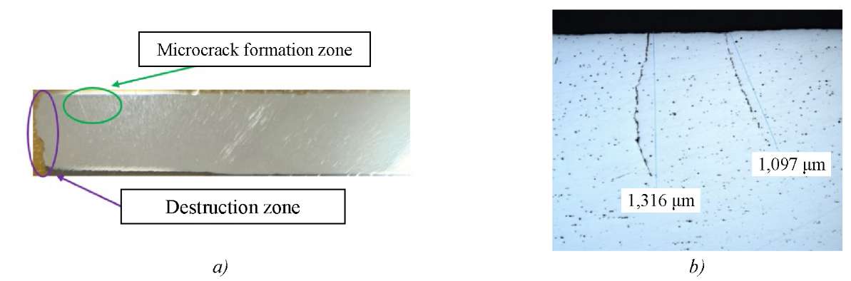
Fig. 7. Macrostructure and microcracks in the cross-section of 09G2S steel sample near the fracture zone after cyclic bending: a — general view of the cross-section with the identification of the fracture area and the area of microcrack formation; b — micrography of microcracks in cross-section indicating the characteristic lengths of 1097 and 1316 µm (×100)
It should be noted that a decrease in fractal dimension D0 on the outer surface was accompanied by similar changes in other multifractal parameters.
The local drop in all three parameters (D0, D1–D0 and f60) was consistent with Hr anomalies detected by magnetic measurements, which confirmed the general nature of damage accumulation and microcrack origin.
To visually confirm the patterns identified by the multifractal and magnetic parameters, the microstructure of the samples in the characteristic zones was analyzed. The images shown in Figure 8 demonstrate a gradual disruption of the structure's order — from the inner part to the outer surface, where microcracks formed.

Fig. 8. Microstructure of parts of the cross-section of the sample wall in the fracture zone:
a — external; b — central; c — internal
On the microstructure of the outer surface adjacent to the fracture zone, there was a high degree of fragmentation of ferrite and perlite grains, as well as local areas with uneven contrast, indicating the development of plastic deformation. Initial microcracks and submicroscopic fractures along the grain boundaries were visible, accompanied by a loss of clarity of their outlines and local misorientation of substructural elements (Fig. 8).
The comparison of microstructures of the outer, central and inner parts of the sample in the fracture zone (Fig. 8) showed a pronounced gradient in the degree of degradation of the structure in thickness. The inner part maintained ordered ferrite-pearlite morphology, clear grain boundaries, and a uniform phase distribution, corresponding to high values of fractal dimension D0 and uniformity parameter f60. The initial restructuring of the grain-subgrain structure was observed in the central zone: small-angle sub-boundaries and local contrasts appeared, indicating an increase in internal instability and a decrease in D1–D0. parameter. Finally, maximum fragmentation and local microcracking occurred on the outer surface, which was accompanied by a decrease in all three fractal indicators: D0, D1–D0 and f60.
This distribution of microstructural features was fully consistent with the results of multifractal analysis and magnetic measurements: the outer part, where the greatest loss of structural complexity occurred, coincided with the area of magnetic anomalies Hr and reflected the zone of maximum damage accumulation. Thus, a comparison of microstructural, fractal and magnetic data confirmed that the degradation of the material had a pronounced gradient character — from a stable internal structure to a destroyed surface, where a focus of fatigue failure was formed.
The resulting set of magnetic and multifractal features was considered as the basis for the subsequent construction of models for assessing the technical condition and predicting the remaining resource. The generated array of parameters can be used in the future as a training base for intelligent technical condition assessment systems, which opens up opportunities for building more stable and adaptive methods of predictive monitoring.
Conclusion. A comprehensive study of 09G2S steel during cyclic bending has shown a uniform pattern of evolution of magnetic, fractal and microstructural parameters reflecting the processes of damage accumulation and destruction. Magnetic measurements of the distribution of the resulting field strength Hr revealed local anomalies coinciding with areas of increased residual stresses and localization of deformation near the outer surface.
The results of the multifractal analysis of micrographs performed in the MFRDrom Fast program confirmed the connection of magnetic anomalies with microstructure degradation. There was a consistent decrease in fractal dimension D0, latent periodicity parameter D1–D0, and degree of uniformity f60, indicating the destruction of the self-similar organization and an increase in the randomness of the substructure.
Metallographic analysis revealed a clear degradation gradient in thickness: the inner part maintained a stable ferrite-pearlite structure, the central part was characterized by partial fragmentation, and the outer part was most destroyed and contained microcracks. A comparison of these three levels showed consistent behavior of the parameters Hr, D0, D1–D0 and f60, which confirmed their interrelated nature.
Thus, the degradation of 09G2S steel during cyclic bending has a multilevel character: magnetic changes reflect the accumulation of defects, fractal parameters reflect the destruction of a large-scale structure, and microstructural analysis is the final stage of microcracking. The combination of these features can be used as a single diagnostic criterion for the condition of the material and the basis for assessing the residual life of elements operating under variable loads.
1. Kadyrov RO, Shermatov DN, Gafarova VA, Kuzeev IR. Changes in the Magnetic Characteristics of a Model Specimen of a Tank’s Utor Assembly as a Function of Accumulated Damage. News of the Tula State University. Technical Sciences. 2025;4:68–79. (In Russ.)
2. Kaappa S, Santa-aho S, Honkanen M, Vippola M, Laurson L. Magnetic Domain Walls Interacting with Dislocations in Micromagnetic Simulations. Communications Materials. 2024;5:256. https://doi.org/10.1038/s43246-024-00697-9
3. Boyao Lyu, Shihua Zhao, Yibo Zhang, Weiwei Wang, Haifeng Du, Jiadong Zang. MagNet: Machine Learning Enhanced Three-Dimensional Magnetic Reconstruction. arXiv. 2022;2210.03066. https://doi.org/10.48550/arXiv.2210.03066
4. Broadway DA, Flaks M, Dubois AEE, Maletinsky P. Reconstruction of Non-Trivial Magnetization Textures from Magnetic Field Images Using Neural Networks. Mesoscale and Nanoscale PhysicsarXiv. 2024;2412.19381. https://doi.org/10.48550/arXiv.2412.19381
5. Ali Tabatabaeian, Ahmad Reza Ghasemi, Mahmood M Shokrieh, Bahareh Marzbanrad, Mohammad Baraheni, Mohammad Fotouhi. Residual Stress in Engineering Materials: A Review. Advanced Engineering Materials. 2021;23(5):1–65. https://doi.org/10.1002/adem.202100786
6. El-Achkar T, Weygand D. Free Surface Acts as Dislocation Sink in Cyclic Loading. In book: Fatigue of Materials at Very High Numbers of Loading Cycles. Springer; 2018. P. 395–416.
7. Polák J. Role of Persistent Slip Bands and Persistent Slip Markings in Fatigue Crack Initiation in Polycrystals. Crystals. 2023;13(2):220. https://doi.org/10.3390/cryst13020220
8. Romanov AE, Kolesnikova AL. Micromechanics of Defects in Functional Materials. Acta Mechanica. 2021;232(5):1901–1915. https://doi.org/10.1007/s00707-020-02872-8
9. Gaur V, Doquet V, Persent E, Mareau C, Roguet É, Kittel J. Surface Versus Internal Fatigue Crack Initiation in Steel: Influence of Mean Stress. International Journal of Fatigue. 2016;82(3):437–448. https://doi.org/10.1016/j.ijfatigue.2015.08.028
10. Mandelbrot BB. The Fractal Geometry of Nature. W.H. Freeman and Company; 2021. 500 p.
11. Fernández R, González‑Doncel G, Garcés G. Fractal Analysis of Strain‑Induced Microstructures in Metals. In book: Fractal Analysis – Selected Examples. IntechOpen; 2020. https://doi.org/10.5772/intechopen.91456
12. Patiño‑Ortiz M, Patiño‑Ortiz J, Martínez‑Cruz MÁ, Esquivel‑Patiño FR, Balankin AS. Morphological Features of Mathematical and Real‑World Fractals: A Survey. Fractal and Fractional. 2024;8(8):440. https://doi.org/10.3390/fractalfract8080440
13. Vstovskii GV, Kolmakov AG, Bunin IZh. Introduction to Multifractal Parameterization of Material Structures. Izhevsk: Scientific Publishing Center “Regular and Chaotic Dynamics”; 2002. 116 p. (In Russ.)
14. Vstovskii GV. Elements of Information Physics. Moscow: MSIU; 2002. 258 p. (In Russ.)
15. Botvina LR, Petersen TB, Tyutin MR. The Acoustic Gap as a Diagnostic Sign of Prefracture. Doklady Physics. 2018;479(5):514–518. (In Russ.) https://doi.org/10.7868/S0869565218110087
16. Shermatov JN, Naumkin EA, Kuzeev IR, Rubtsov AV. Change of Ultrasonic Waves Speed Spreading in a Material of the Reaction Furnace Coil during Operation Process. Petroleum Engineering. 2019;17(5):81–88. (In Russ.) https://doi.org/10.17122/ngdelo-2019-5-81-88
17. Ailin Li, Wenwu Zhong, Cong Yu, Xin Zhang, Tao Li, Zheng Fei. Study on Rock Damage Mechanics in the Sustainable Development of the Red Sandstone Area in China: Taking Zhongjiang County as an Example. Frontiers in Earth Science. 2025;12:1484633. https://doi.org/10.3389/feart.2024.1484633
Dzhamshеd N. Shermatov, Cand. Sci. (Eng.), Associate Professor of the Department of Technological Machines and Equipment
1, Kosmonavtov St., Ufa, 450064
Scopus ID: 58073438400
Alexander O. Borisov, Postgraduate Student of the Department of Technological Machines and Equipment
1, Kosmonavtov St., Ufa, 450064
ResearcherIDNVM-5431-2025
Victoria A. Gafarova, Cand. Sci. (Eng.), Associate Professor of the Department of Technological Machines and Equipment
1, Kosmonavtov St., Ufa, 450064
Scopus ID: 57151391500
ResearcherIDC-9969-2017
Iskander R. Kuzeev, Dr. Sci. (Eng.), Professor of the Department of Technological Machines and Equipment
1, Kosmonavtov St., Ufa, 450064
The research is focused on developing a method for the early detection of fatigue failure in metal structures by analyzing changes in the magnetic field of the material. A novel approach based on the spatial registration of the strength of the permanent magnetic field at various points along a steel sample under cyclic loading has been proposed. Experimental results have shown that abnormal changes in the magnetic signal during the first forty to fifty percent of operation time indicate the formation of areas prone to destruction with high probability. This method demonstrates high sensitivity to early stages of material deterioration and allows for the localization of potential damage areas before visible cracks occur.
Shermatov D.N., Borisov A.O., Gafarova V.A., Kuzeev I.R. Local Gradient Indicator of Magnetic Variability under Cyclic Loading of Steels. Safety of Technogenic and Natural Systems. 2026;10(1):47-60. https://doi.org/10.23947/2541-9129-2026-10-1-47-60. EDN: BZBQLB


 Email this article (Login required)
Email the author (Login required)
Email this article (Login required)
Email the author (Login required) Safety of Technogenic and Natural Systems
eISSN 2541-9129
Contact with: Publisher / Editorial Office of the Journal
Publisher: Don State Technical University - DSTU, Rostov-on-Don, Russia - https://donstu.ru/en/
Editor-in-Chief: Meskhi Besarion Chokhoevich, Doctor of Technical Sciences, Professor (Rostov-on-Don, Russia)
Don State Technical University
1, Gagarin Sq., Rostov-on-Don, 344003, Russia
tel.: +7 (863) 2738-372, e-mail: btps@donstu.ru
1, Gagarin Square, Rostov-on-Don, 344003, Russian Federation, Don State Technical University
Processing of personal data


